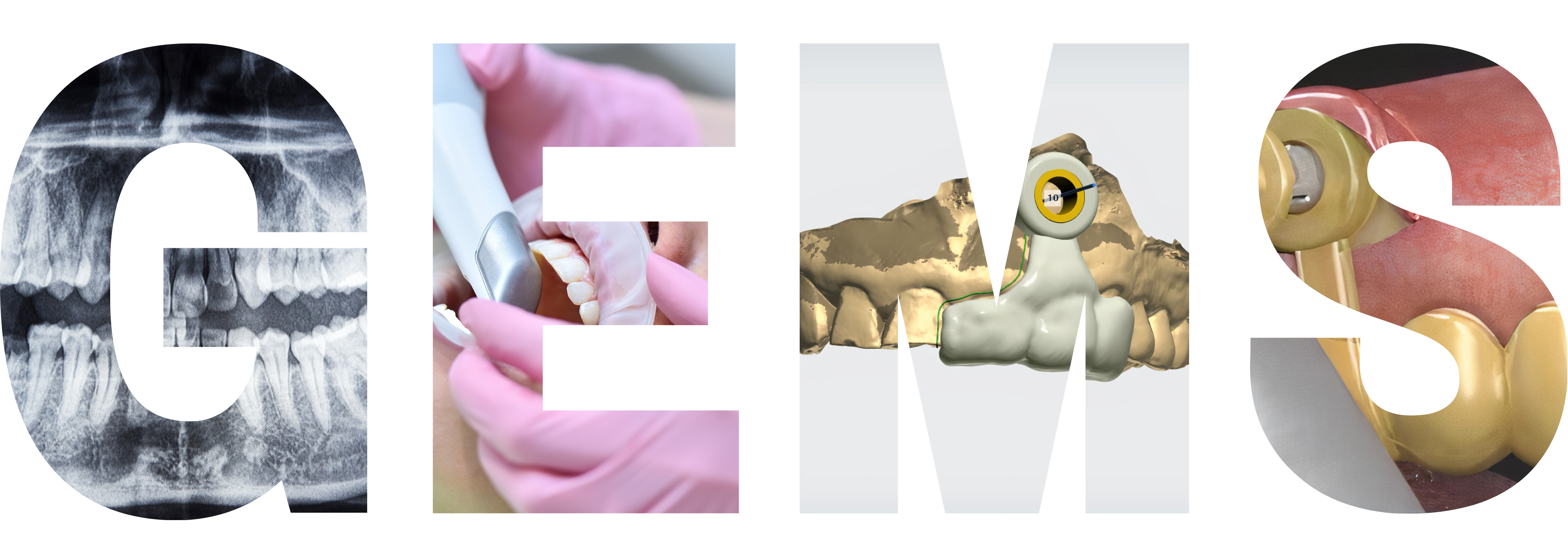
Guided Endodontics Microsurgery (GEMS) is a relatively new technique for endodontic microsurgery that is rapidly gaining popularity. It is a minimally invasive surgical method that is guided by a 3D-printed template and aims to improve the predictability and precision of endodontic microsurgery.
The GEMS technique involves the use of a cone-beam computed tomography (CBCT) scan to create a 3D image of the root canal system and the surrounding bone. From this image, a 3D-printed template is designed that guides the endodontic surgeon during the surgery. The template is customized for each patient’s unique anatomy and takes into account the size and shape of the root canal system, as well as the position of the adjacent teeth and vital structures.
During the surgery, the template is positioned over the tooth, and the surgeon follows the preplanned path to access the root canal system. This helps to reduce the risk of damaging surrounding structures and ensures that the surgery is carried out with the utmost precision. The template also allows for more accurate suturing and closure of the surgical site, which can promote better healing and reduce the risk of complications.
The GEMS technique has several advantages over traditional endodontic microsurgery methods. It allows for a more minimally invasive approach, reducing trauma to the patient and promoting faster healing times. It also improves the predictability and accuracy of the surgery, reducing the risk of complications and improving the overall success rate.
Although GEMS is a relatively new technique, it has shown promising results in clinical studies. It has been shown to be effective in treating periapical lesions and achieving successful outcomes in cases that were previously considered difficult or impossible to treat. As such, it is rapidly becoming an essential tool in the field of endodontic microsurgery and is poised to revolutionize the way that these procedures are carried out.
GEMS workflow process involves the use of advanced technology to design a customized 3D-printed template that guides the endodontic surgeon during the surgery. This results in improved accuracy, predictability, and success rates compared to traditional endodontic microsurgery methods.
Customized 3D-printed templates: The use of customized 3D-printed templates in GEMS is a significant innovation that improves the accuracy and predictability of endodontic microsurgery.
Minimally invasive approach: The GEMS technique involves a more minimally invasive approach compared to traditional methods, resulting in reduced trauma to the patient and promoting faster healing times.
Improved accuracy and success rates: The use of a 3D-printed template in GEMS ensures that the surgery is carried out with the utmost precision, reducing the risk of damaging surrounding structures. This improved accuracy and predictability have been shown to result in higher success rates in clinical studies.
Advanced technology: GEMS utilizes advanced technology, such as cone-beam computed tomography (CBCT) scans and 3D printing, to design customized templates that guide the surgeon during surgery.
Faster healing times: Due to its minimally invasive approach, GEMS promotes faster healing times and reduces the risk of complications.
Increased safety: The use of a 3D-printed template in GEMS reduces the risk of damaging surrounding structures and improves overall safety during endodontic microsurgery.
Overall, GEMS is an innovative technique that utilizes advanced technology and a more minimally invasive approach to improve the accuracy, predictability, and success rates of endodontic microsurgery.
The Guided Endodontics Microsurgery (GEMS) technique has several advantages over traditional endodontic microsurgery methods:
Minimally invasive approach: GEMS involves a more minimally invasive approach compared to traditional methods, resulting in reduced trauma to the patient and promoting faster healing times.
Improved accuracy: The use of a 3D-printed template in GEMS ensures that the surgery is carried out with the utmost precision, reducing the risk of damaging surrounding structures.
Predictability: GEMS improves the predictability of endodontic microsurgery, allowing the surgeon to plan and execute the procedure with greater accuracy.
Customization: The GEMS technique utilizes customized 3D-printed templates that are tailored to each patient’s unique anatomy, improving the accuracy of the surgery.
Faster healing: Due to its minimally invasive approach, GEMS promotes faster healing times and reduces the risk of complications.
Higher success rates: The improved accuracy and predictability of the GEMS technique have been shown to result in higher success rates in clinical studies, making it a valuable tool in endodontic microsurgery.
Overall, GEMS has several advantages over traditional endodontic microsurgery methods, making it a promising and innovative technique in the field of endodontics.

GEMS Kit
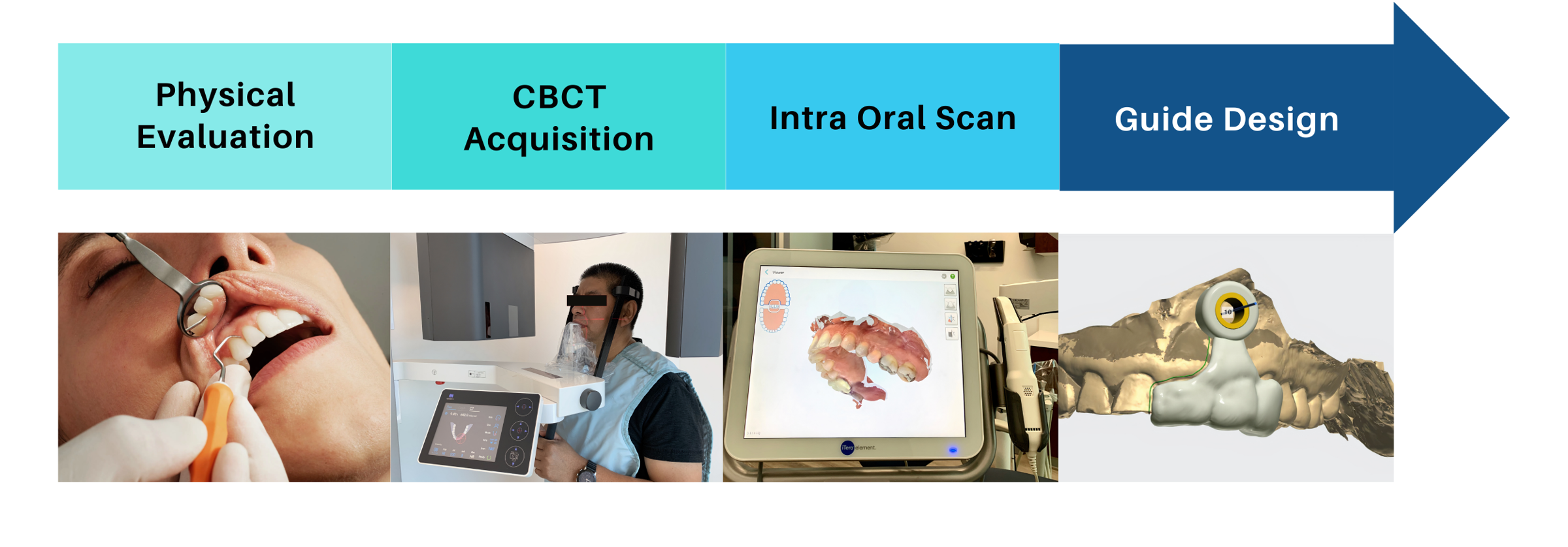
The Guided Endodontics Microsurgery (GEMS) workflow process involves several steps:
Cone-beam computed tomography (CBCT) scan: The first step in the GEMS workflow process is to perform a CBCT scan of the affected tooth and surrounding structures. This scan is used to create a 3D image of the root canal system and surrounding bone.
3D-printed template design: Using specialized software, the 3D image is used to design a customized 3D-printed template that will guide the endodontic surgeon during the surgery. The template takes into account the size and shape of the root canal system, as well as the position of the adjacent teeth and vital structures.
Surgical preparation: Prior to the surgery, the patient is prepared in the usual manner for endodontic microsurgery. This may involve local anesthesia and other preparations as necessary.
Template placement: The 3D-printed template is then placed over the tooth, and the surgeon follows the preplanned path to access the root canal system. This helps to reduce the risk of damaging surrounding structures and ensures that the surgery is carried out with the utmost precision.
Surgery: Once the template is in place, the surgeon carries out the endodontic microsurgery using traditional techniques. The surgeon uses the template as a guide to access the root canal system and perform the necessary procedures.
Suturing and closure: After the surgery is complete, the surgical site is closed using the 3D-printed template to ensure accurate suturing and closure of the surgical site. This promotes better healing and reduces the risk of complications.